Your AI Passed Testing — But on Whose Data? An Insider's Guide to Evaluation That Actually Works
I've sat inside the AI training pipeline. I've reviewed the datasets. I've seen what companies call "validated" and watched the same models collapse the moment they encounter a real pediatric case. The evaluation problem in radiology AI is not theoretical — it is structural, and it starts with the data.
If you're building an AI tool for medical imaging and you think your evaluation is solid, this article is for you. Not because your team is careless — but because the gaps I'm about to describe are industry-wide, and most companies don't realize they exist until the tool is already in clinical use.
Evaluation Starts After Training — But It Depends on What Happened Before
The evaluation phase is where you answer the most important question about your AI: How well does this tool actually work? Not on your curated benchmark. Not in your demo. In the real world, on real patients, with the kind of case mix a hospital sees every day.
But meaningful evaluation is impossible without meaningful data. And this is where most pipelines break down — not because companies skip evaluation, but because the datasets they use for it are flawed from the start.
Robust Datasets: The Foundation Everything Else Stands On
A robust evaluation dataset has all kinds of case mixes — the common and the rare, the textbook presentations and the atypical ones, the easy calls and the cases that make experienced radiologists pause.
In pediatric imaging, this is especially critical because:
- Normal anatomy changes with age. A chest X-ray of a 3-month-old looks fundamentally different from that of a 12-year-old. A dataset that skews toward one age bracket will produce evaluation metrics that are meaningless for the others.
- Disease prevalence varies by age. Neuroblastoma peaks in infants. Osteosarcoma peaks in adolescents. Intussusception clusters between 6 months and 3 years. A dataset that doesn't capture this distribution will produce an AI that looks great on paper and fails in practice.
- Imaging protocols differ across institutions. Dose settings, contrast protocols, and acquisition techniques vary. A robust dataset reflects this variability — because your model will encounter it in deployment.
- Rare conditions matter. A model that detects common fractures with 98% accuracy but misses Salter-Harris Type V fractures entirely is not a safe model for pediatric care. Your evaluation data must include the long tail of pathology, not just the top 10 diagnoses.
The insider take: I've reviewed datasets from companies building pediatric AI tools where the entire "pediatric" set was actually adolescents aged 14-17 — cases that look nearly identical to young adults. The model appeared to work. It didn't work on the patients who needed it most: neonates, infants, and young children.
The Triple-Dataset Rule: Training, Validation, and Testing
This is fundamental to any machine learning pipeline, but I still see it violated — especially when pediatric data is scarce and teams are tempted to cut corners.
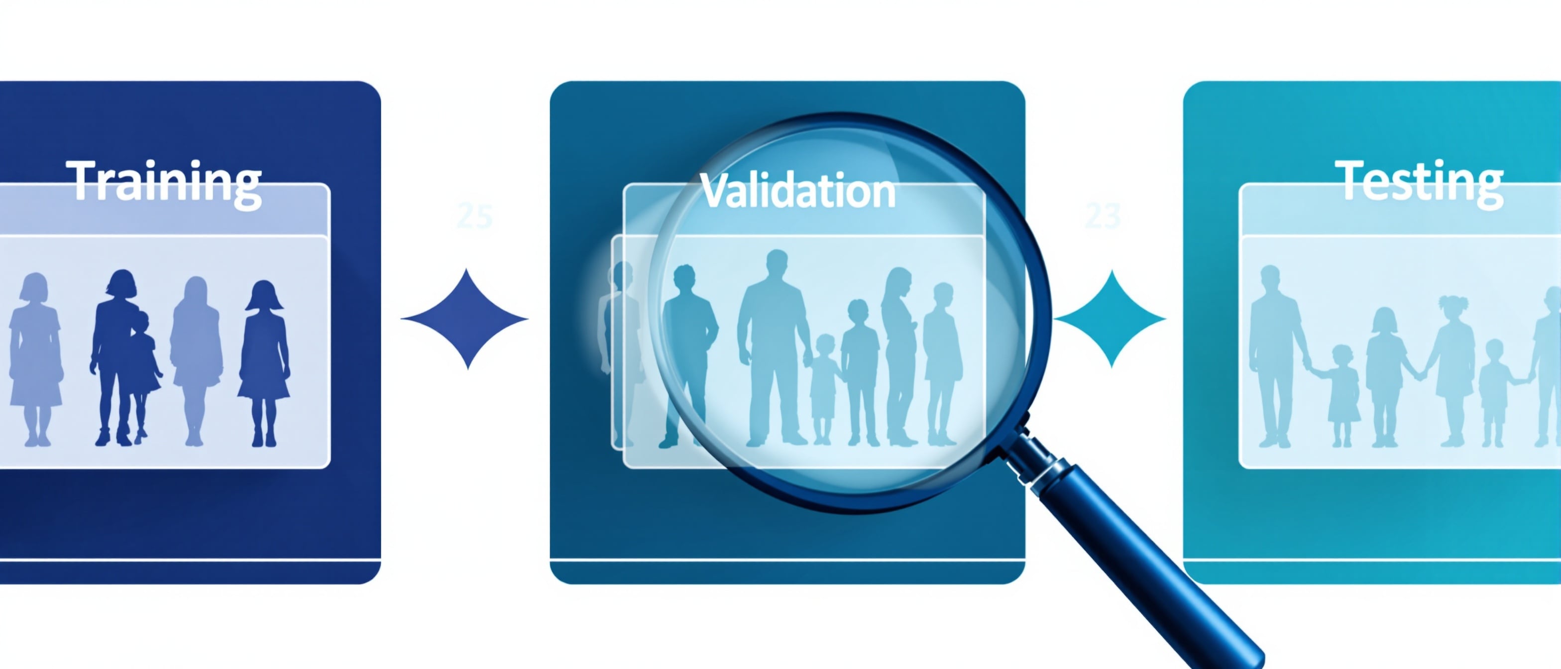
Every AI tool needs three independent datasets:
- Training data — what the model learns from. This is the largest set, and it shapes the model's internal representations.
- Validation data — what you use to tune the model during development. Hyperparameter selection, architecture decisions, and early stopping criteria all depend on this set.
- Testing data — the final, held-out dataset that the model has never seen. This is your honest measure of real-world performance.
The reason you need all three is overfitting — the tendency for a model to memorize the training data rather than learn generalizable patterns. A model that has overfit will show impressive metrics on familiar data and deteriorate on anything new.
The validation set guards against this during development. The test set confirms it after development is complete. Skip either one, and you are flying blind.
The Critical Rule: Independence
These three datasets must be independent. No overlap. No shared patients. No images from the same study appearing in different sets. If there is any contamination between them — any leakage — your evaluation metrics are meaningless.
Data leakage is one of the most common and most devastating errors in medical AI. It comes in several forms:
- Patient leakage: The same patient's images appear in both training and testing sets. The model recognizes the patient, not the pathology. This is especially common in longitudinal studies where a child has multiple imaging exams over time.
- Institutional leakage: All data comes from one hospital. The model learns that institution's specific imaging characteristics (scanner artifacts, protocol quirks, annotation style) rather than actual clinical findings. Performance collapses at any other site.
- Temporal leakage: Training and testing data come from the same time period. Seasonal disease patterns, staffing changes, and equipment upgrades can create artificial correlations that inflate performance.
The insider take: I've seen AI papers reporting 95%+ accuracy on pediatric imaging tasks where the training and testing sets both came from the same 200 patients at a single institution. Remove the leakage, test on an external dataset, and the accuracy drops to 60-70%. The model wasn't detecting disease — it was detecting the hospital.
Bias: The Silent Threat to Your AI's Credibility
Even with perfectly separated datasets and rigorous evaluation protocols, bias can undermine everything. In pediatric imaging AI, two forms of bias are particularly dangerous — and particularly common.
Single-Rater Bias
Most AI training data is labeled by a single annotator. One radiologist reviews the case, assigns a label, and that label becomes ground truth. But radiology is not always black and white. There are judgment calls, borderline findings, and legitimate disagreements between experienced readers.
When a single rater labels your data:
- The model learns that rater's specific biases, thresholds, and blind spots
- Borderline cases are resolved by one person's judgment rather than clinical consensus
- Subtle findings that one reader might miss become systematically absent from the training data
- Inter-reader variability — a known factor in radiology — is ignored entirely
The solution is consensus rating: multiple qualified readers independently review each case, and disagreements are resolved through structured adjudication. This is more expensive and more time-consuming. It is also the only way to produce labels that are clinically defensible.
In pediatric radiology, this is even more critical. The subspecialty is so narrow that subtle findings — a small cortical irregularity that could be a buckle fracture or a normal variant, a mildly prominent thymus versus early lymphoma — require genuine pediatric radiology expertise to adjudicate. A consensus panel of general radiologists is not equivalent to a consensus panel of fellowship-trained pediatric radiologists.
Population Bias
A model is only as fair as the population it was trained on. If your training data over-represents certain demographic groups and under-represents others, your model will perform unevenly — and the patients who are under-represented will bear the consequences.
In pediatric imaging AI, population bias shows up in several dimensions:
- Age group bias: Models trained predominantly on older children may fail on neonates and infants, where the anatomy and pathology are most different from adults.
- Racial and ethnic bias: Normal anatomical measurements, bone density, and disease prevalence vary across racial and ethnic groups. A model trained on a non-representative population may systematically misdiagnose or miss findings in under-represented groups.
- Institutional bias: A dataset from one tertiary referral center reflects that center's specific patient population — typically sicker, more complex cases. This creates a model that may over-detect in community hospital settings and under-detect in lower-acuity populations.
- Sex-based bias: Some conditions are more prevalent in boys versus girls (or vice versa). If your data doesn't reflect these distributions, the model inherits an imbalanced understanding of disease presentation.
The insider take: A fracture detection AI that works well on Caucasian adolescents may have never been exposed to the normal skeletal variants common in children of African descent — and may flag normal anatomy as pathological. This isn't hypothetical. It's happening now, and without diverse evaluation datasets, no one catches it until it reaches the patient.
What Rigorous Evaluation Actually Looks Like
If you are serious about deploying AI in pediatric imaging, your evaluation framework should include:
- Age-stratified performance analysis — not a single accuracy number, but performance broken down by neonate, infant, toddler, child, and adolescent cohorts
- Multi-site testing — external validation on data from institutions your model has never seen
- Consensus-labeled ground truth — annotations from multiple fellowship-trained pediatric radiologists with structured disagreement resolution
- Bias auditing — systematic analysis of performance across racial, ethnic, sex, and age subgroups
- Failure mode analysis — not just where the model succeeds, but where and how it fails, and whether those failures cluster in vulnerable populations
- Clinical significance assessment — moving beyond sensitivity and specificity to ask: Does this error matter clinically? Would it change management? Could it harm a patient?
This level of evaluation requires subspecialty expertise at every step. Not just to read the images, but to design the evaluation framework itself — to know which case mixes matter, which edge cases to include, which failure modes are clinically dangerous versus clinically irrelevant.
Where NeuraTots Fits In
This is what we do. We have been inside the pipeline. We have seen the gaps. And we bring the one thing that cannot be automated or approximated: fellowship-trained pediatric radiology expertise applied systematically to AI evaluation.
Our evaluation and validation services include:
- Robust dataset design — we help you build evaluation datasets with appropriate case mixes, age stratification, and demographic diversity
- Consensus annotation — multi-reader panels of fellowship-trained pediatric radiologists, not general radiologists, eliminating single-rater bias
- Independent dataset curation — we ensure your training, validation, and testing sets are truly independent, with no patient or institutional leakage
- Bias detection and mitigation — systematic auditing across population subgroups before your model reaches clinical deployment
- Regulatory-grade documentation — evaluation frameworks that satisfy FDA and CE marking requirements for pediatric medical device submissions
Your AI may have passed testing. The question is: whose data was it tested on? If the answer doesn't include a robust, independent, consensus-labeled pediatric dataset with full demographic representation — the evaluation isn't complete.
We can help you get there.

Dr. Rotimi Komolafe
Founder & Principal Consultant, NeuraTots
North American fellowship-trained pediatric radiologist. Published AI researcher and expert AI/Radiomics peer reviewer for the Journal of Pediatric Radiology.